
Revise
Microscopy
Microscopy
Cells are too small to see with the human eye, but thanks to the development of technology, we are able to view cells and even their subcellular structures under microscopes.
Microscopes
Light microscopes use light and lenses to create a magnified image of a specimen. Their development enabled scientists to view individual cells and their larger subcellular structures such as nuclei. Light microscopes have been further developed, with improved magnification and resolution, but typically have a maximum magnification of only \times 1500 and resolutions of up to 0.2\text{ μm}. This means that the amount of detail that can be seen via a light microscope is limited.
Electron microscopes have much greater magnification and resolving power because they use electron beams instead of light which have a much smaller wavelength. The development of the electron microscope allowed scientists to see cells in much more detail including the internal structures of mitochondria, chloroplasts and nuclei, and tiny structures like ribosomes and plasmids.

Note:
‘Resolution’ or ‘Resolving power’ is just the ability to distinguish between two points. You can think of resolution in the context of a photograph – if you enlarge the image, after a certain point you will not be able to see any more detail and it just becomes more blurry.

Magnification
The magnification of an image can be calculated by a simple formula:
\text{Magnification} = \dfrac {\text{Size of the image}}{\text{Actual size of object}}
This formula can be rearranged to find the size of an image or an object.
An easy way to do this is by using this formula triangle:

Just cover up the part of the triangle you want to find and follow the equation that is left behind.
Example: A student measures an image of a cell using a ruler. It is 55\text{ mm} wide and the image has been magnified by a factor of \times 5000. What is the actual width of the cell in \text{μm}?
Use the formula triangle to work out what calculation to do. Cover up the ‘actual size’ portion of the triangle and notice that what remains is image size divided by magnification.
\text{Actual size =}\dfrac{\text{Image size}}{\text{Magnification}}
Substitute the numbers from the question into the equation:
\text{Actual size} =\dfrac{55}{5000} =0.011\text{ mm}
To convert the answer into μm you need to times by 1000 because there are 1000 μm in a mm (more on this in other topics).
0.011\times 1000 = 11 \text{ μm}
Standard Form
Questions may also ask you to give an answer in standard form. This is just an easier way of writing a number when it is really big or really small and so has lots of zeros in it.
In order to convert to standard form you move the decimal point, left or right, until it is a number between 1 and 10. The number of places the decimal point has moved is represented by a power of 10. If the decimal point moves left the the power will be positive and if it moves to the right it will be negative.
Example:
0.00023 in standard form would be 2.3\times 10^{-4} as get to 2.3 the decimal point moves 4 places to the right.
46000000 in standard form would be 4.6\times 10^{7} as to get to 4.6 the decimal point moves 7 places to the left.
Required Practical
Using a light microscope to draw and label a cell with a magnification scale.
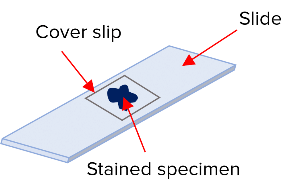
Prepare the slide
- Add a drop of water to a clean slide.
- Carefully extract the cells of interest and place them on the slide, in the water – common choices are human cheek cells (animal) and onion epidermal cells (plant).
- Highlight the cells using an appropriate stain (Iodine for onion cells and methylene blue for cheek cells).
- Finally, place a cover slip over the top of the specimen.
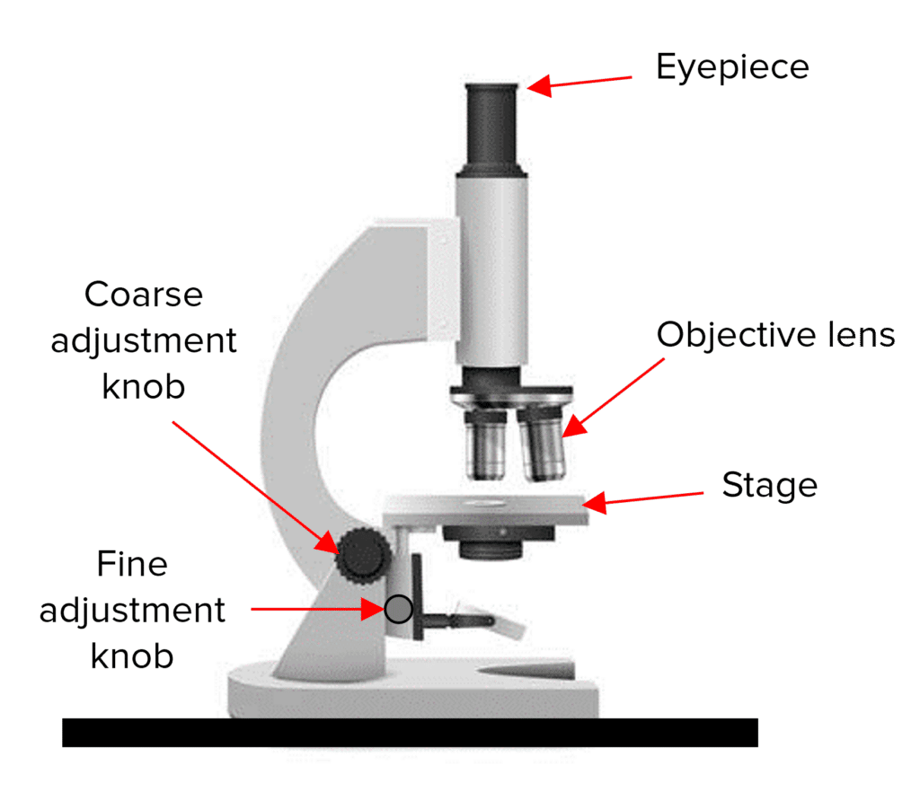
View slide under the light microscope
- Carefully place the slide onto the stage and clip it in place.
- Select the objective lens with the lowest power and therefore lowest magnification.
- While looking down the eyepiece, move the stage up and down using the coarse adjustment knob until the image becomes more focussed.
- Use the fine-adjustment knob to further focus the image until it is clear.
- Switch to a higher powered objective lens and refocus if greater magnification is required.
Drawing your findings
- Biological drawings should be scientific so draw what you see down the microscope with clear unbroken lines and in the correct proportions.
- Label the different cell structures with clear straight lines, add a title and state the magnification that the cell was observed under.
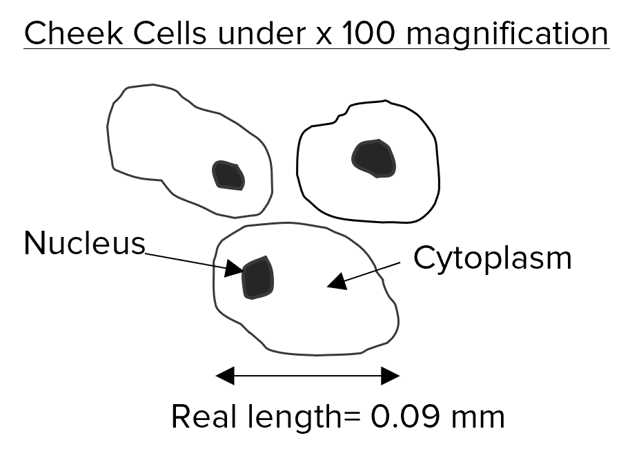
Create and add a scale bar to the drawing
- Clip an eyepiece graticule to the top of the slide and select the \times 100 objective lens on the microscope.
- Line the cells up and count how many fit along 1\text{ mm} on the eyepiece graticule.
- Divide 1 (\text{mm}) by the number of cells counted to find the length of one cell and add this to the drawing.
Microscopy Example Questions
Question 1: Name an advantage of using an electron microscope over a light microscope.
[1 mark]
Electron microscopes have much greater magnification and resolving power so smaller subcellular structures can be viewed in great detail and studied e.g. ribosomes, plasmids and the internal structures of mitochondria, chloroplasts and nuclei.

Question 2: A specimen is 38 \text{ μm} long. It is viewed under a microscope with a magnification of \times 100. Calculate the length of the image produced in \text{mm}.
[2 marks]
\text{Image size} = 100 \times 38= 3800\text{ μm}
3800\text{ μm} = 3.8 \text{ mm}
Question 3: Describe how a student would prepare a slide of onion cells, ready to be viewed under a light microscope.
[3 marks]
Add a drop of water to a slide then carefully extract the onion cells and place them in the water on the slide. Then stain the cells with iodine to make them more visible and place a cover slip over the top.
Specification Points Covered
AQA GCSE –
4.1.1.2 Animal and plant cells
4.1.1.5 Microscopy
Microscopy Worksheet and Example Questions
Microscopy Questions
GCSEOfficial MME